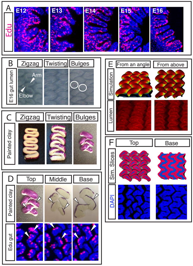
Figure 5. The formation of villi from zigzags involves non-uniform proliferation and a complex change in topography.
(A) Transverse sections of guts labeled for 4 hours with Edu in ovo (red) guts show patterns of proliferation over time. (B) Luminal views of guts from E15 to E16 as villi form. The “arm” of the zigzag rotates at the “elbow”, the circles denote the resulting pockets of mesenchyme surrounded by endoderm that will each become a villus. (C) Clay models, purple label represents proliferating regions. Clay model is twisted to mimic change in topography seen in B. (D) Top - Labeled, twisted model of E16 gut is sliced with a razor blade to reveal label localization. Bottom - Edu label in longitudinal sections of E16 guts, arrowheads highlight similarity of pattern. (E) Top - a simulation that incorporates non-uniform proliferation along with measured geometrical and biophysical parameters show villi morphogenesis. Bottom – corresponding images of the chick lumen (red color and stained puncta are due to antibody stain and should be disregarded) (F) Top - sections of the simulations in D. Bottom – corresponding sections in chick.

