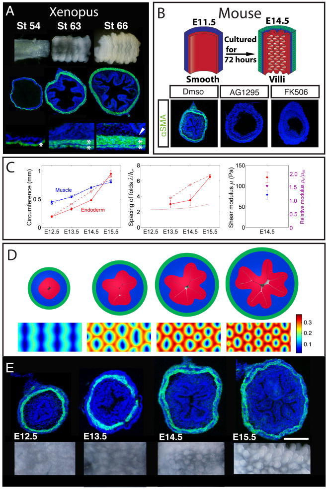
Figure 6. The physical mechanism of villification can be extended to other species.
(A) Luminal pattern formation in Xenopus. Sections labeled with DAPI (blue) and SMA (green). Here the circumferential and outer longitudinal layer form (asterisks) but the inner longitudinal layer does not form (arrowhead) (B) Transverse sections of E11.5 mouse guts (labeled as in A) cultured in vehicle alone (DMSO) or with either 10μm AG1295 or 10μm FK506 for 72 hours, experiment schematized above. (C) Left - circumference of the inner boundary of the muscle and endoderm. Middle - spacing of folds measured along the endoderm and scaled by its thickness. Dotted line is the stress-free thickness of the endoderm-mesenchyme composite. Right - shear moduli of mesenchyme and endoderm, and their ratio. In all panels solid lines correspond to experimental observations and dashed lines to simulations. (D) Cross-sectional (top) and luminal (bottom) images from a simulation based on measurements from the developing mouse gut. Color shows distance of the luminal surface to the center line, relative to the diameter of the tube. (E) Transverse sections (labeled as in A) and whole mount images of the lumen for corresponding stages during mouse villi formation. (n > 3 for culture experiments, error bars represent one SD. Scale bars = 100μm)

