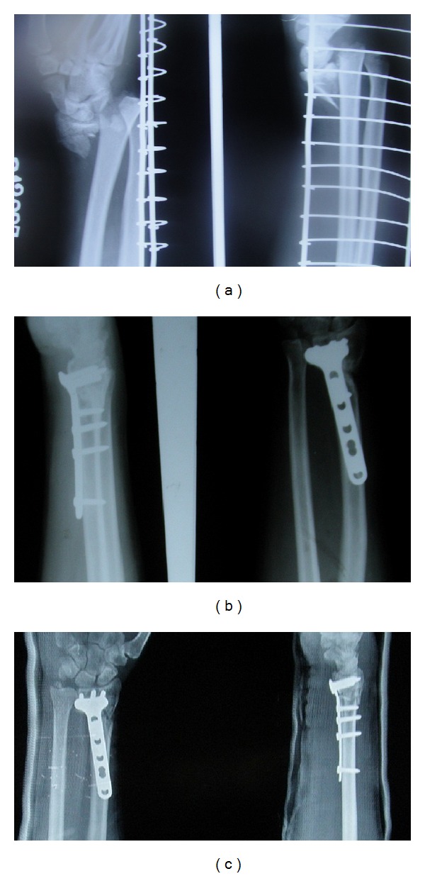
Figure 1.

(a) Preoperative radiographs of an AO type A3 fracture of the distal end of the radius in a 53-year-old male. (b) Postoperative radiographs showing adequate reduction. (c) Radiographs at the final followup, showing that the fracture has united and the radiological parameters are maintained.
