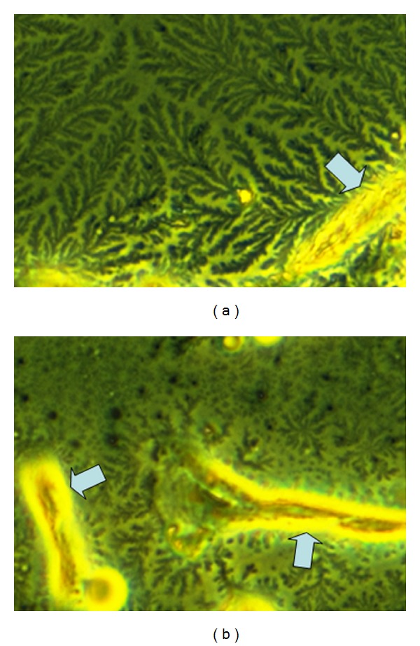
Figure 4.

Illustration of digestion of mesangial cell-related extracellular matrix with hyaluronidase. Mesangial cells were incubated without (a) and with (b) hyaluronidase. Branch-like structures on the cell surface (a) are at least containing hyaluronan, since they are cleaved by hyaluronidase (b) to minor fragments. While high molecular hyaluronan binds water to form a jelly, low molecular hyaluronan forms a liquid solution. So the branches in (a) illustrate the jelly barrier on the cell surface. (a) Mesangial cells form branch-like structures around the outer aspect of the cells (arrow). (b) Hyaluronidase digestion leads to rapid fragmentation of this matrix. Original magnification ×400.
