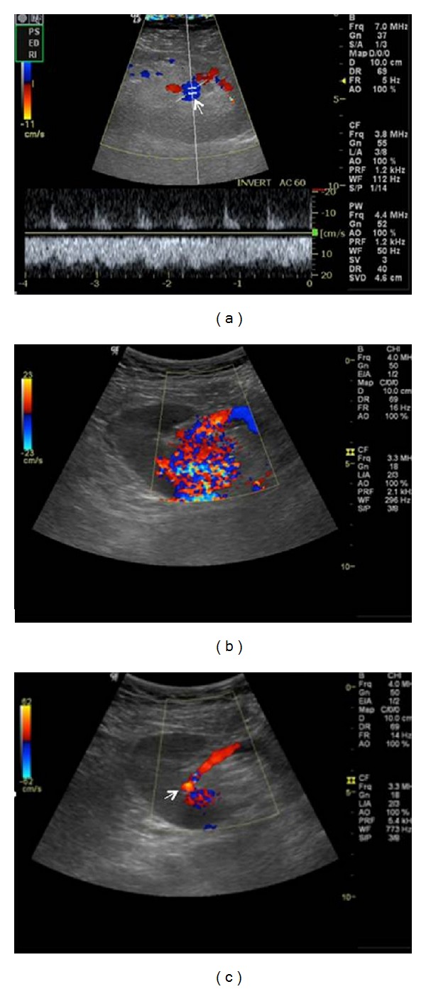
Figure 7.

Intrarenal arteriovenous fistula following biopsy. Color Doppler ultrasound demonstrates a highly vascular lesion (arrow) with aliasing. Spectral Doppler image shows the characteristic mixed arterial venous waveform, with high velocities and low impedence.
