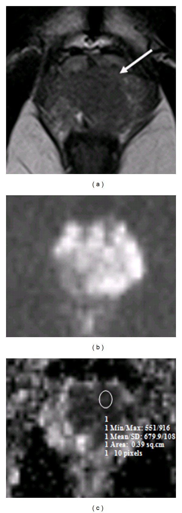
Figure 3.

Patient with high-risk prostate cancer. Pelvic T2W transverse image (a) shows a well-defined low signal intensity region in the peripheral zone on the left base (arrow). (b) DWI; (c) an ROI drawn on ADC map shows restricted diffusion in corresponding left base PZ, scored as Gleason 9 on sextant biopsy.
