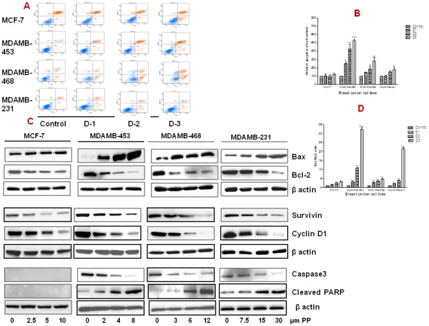
Figure 3. Panepoxydone induces apoptosis in breast cancer cells.

(A) MCF-7, MDA-MB-231, MDA-MB-468 and MDA-MB-453 breast cancer cells were grown in a 6-well plate (1×10⁶ cells/well) and treated with increasing concentrations of PP or DMSO (0.2% vehicle control) for 24 hrs. After treatment, cells were stained with 7-AAD and PE Annexin V followed by flow cytometry. Unstained DMSO-treated cells served as a negative control. Data show a dose-dependent increase in the number of apoptotic cells in all breast cancer cells after treatment with PP as compared to control cells. A representative picture of two independent experiments is shown. (B) Histogram representation of increased number of apoptotic cells in breast cancer cells after treatment with PP. (C) Total protein was isolated from control and PP-treated breast cancer cells and subjected to immunoblotting of apoptosis an dsurvival related proteins. Membranes were stripped and re-probed with anti-actin antibody to ensure equal protein loading. Bax and cleaved PARP was upregulated and Bcl-2, survivin, cyclin D1 and caspase 3 was down-regulated in all breast cancer cells in a dose-dependent manner. (D) Bar diagram indicate the increased Bax/Bcl-2 ratio in all breast cancer cells after PP treatment indicating its role in apoptosis. * indicates statistically significant difference between PP treated and untreated cells at p<0.05 (*), p<0.01(**), and, p<.001(***) levels by student's t-test. Altogether this data indicate the possible role of PP in modulation of apoptosis related genes. Immunoblotting for each protein was performed at least twice.
