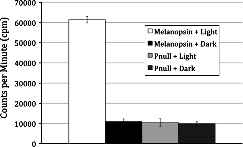
Fig. 2.
Light-dependent phosphorylation of melanopsin in vitro. Membrane preparations containing ~250 pmol of mouse melanopsin were resuspended in kinase buffer after reconstitution with 50 μM 11-ci-retinal (opsin + chromophore). Samples were either exposed to white light for 30 min, or left in the dark. Incorporation of 32P as measured by a filter-binding assay and scintillation counting of WT melanopsin or phospho-null mutant melanopsin. Data represents the average of three measurements; error bars represent standard deviation

