
Figure 6. MWF maps of another healthy 3D T2prep adiabatic spiral brain scan.
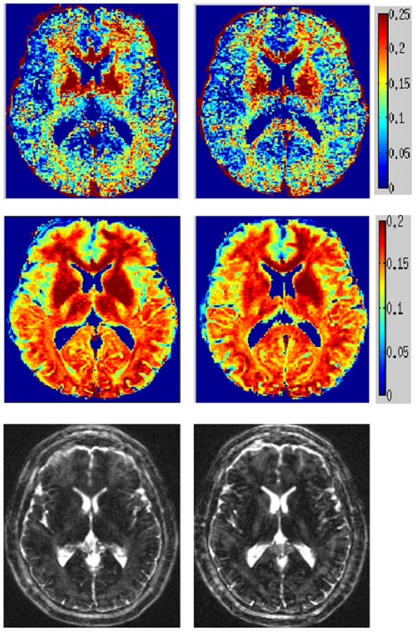
Another healthy brain 3D T2prep adiabatic spiral example, with MWF (top) and T2-weighted image (bottom) of two adjacent axial slices.
Official websites use .gov
A
.gov website belongs to an official
government organization in the United States.
Secure .gov websites use HTTPS
A lock (
) or https:// means you've safely
connected to the .gov website. Share sensitive
information only on official, secure websites.
Another healthy brain 3D T2prep adiabatic spiral example, with MWF (top) and T2-weighted image (bottom) of two adjacent axial slices.