
Figure 8. Illustration of demyelinating lesion in MWF map of a MS patient.
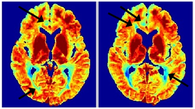
Two axial slices of proposed MWF map of a MS patient. Note the excellent depiction of demyelinating lesions (arrows) and improved definition of callosal and peripheral white matter.