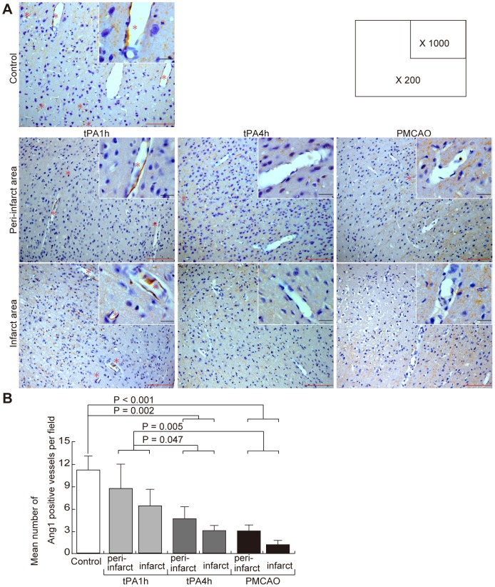
Figure 3. Changes in Ang1-positive vessels due to focal cerebral ischemia.
(A) Immunohistochemical staining with an anti-Ang1 antibody. Representative findings are shown of the peri-infarct and infarct areas of the control, tPA-1h, tPA-4h, and PMCAO groups. High magnification (1,000×) is shown in the upper right of the low magnification (200×) photograph. Ang1-positive vessels were shown by asterisk. tPA, tissue plasminogen activator; PMCAO, permanent middle cerebral artery occlusion; Ang1, angiopoietin-1. The black scale bar is 10 µm, and the red scale bar is 100 µm. (B) Mean number of Ang1-positive vessels. Three locations were chosen randomly in the control cerebral cortex and in each infarct area and peri-infarct area. The figures are the mean number of Ang1-positive vessels from 3 random fields of view of an optical microscope at 200× magnification. Each group N = 5.

