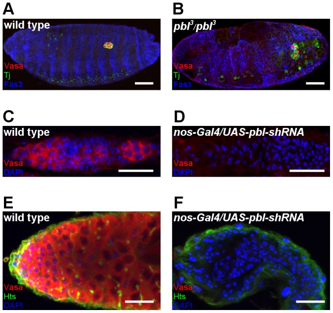
Figure 3. Pbl affects germ-cell development.

(A,B) Immunostaining of a wild type (A) and a homozygous pbl3 mutant embryo (B). Pbl mutants have fewer germ cells, misguided germ cells and abnormally compacted gonads. Vasa staining labels germ cells (red), Tj staining labels the somatic gonad precursor cells (green). Week Tj expression is also detectable in the cenrtral nervous system. The outline of the embryos is marked by Fas3 staining (blue). Scale bar represents 50 µm. (C–F) Immunofluorescence images of adult gonads. Wild type ovariole (C) and testis (E) contain high number of developing germ cells. Nos-Gal4-VP16/UAS-pbl-shRNA (TRiP.GL01092) ovariole (D) and testis (F) lack germ cells. Vasa staining labels germ cells (red), DAPI labels the nuclei (blue). Anterior is to the left. Scale bar represents 20 µm.
