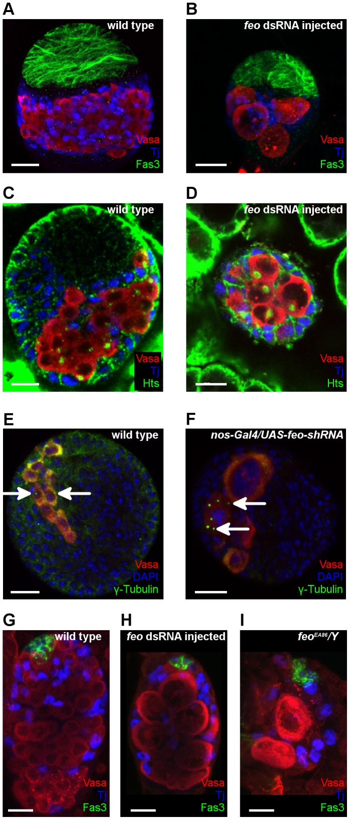
Figure 4. Feo is required for mitosis of larval germ cells.

(A–D) Immunofluorescence images of third stage larval ovaries. Ovaries of larvae injected with feo dsRNA are rudimentary and contain fewer but larger germ cells than the wild type suggesting that the PGCs were unable to undergo mitotic divisions. All ovaries are shown with anterior to the top. Scale bar represents 20 µm. Vasa staining labels the germ cells (red), Tj staining labels the somatic intermingled cells (blue), Fas3 staining labels the anterior somatic cells in (A,B), Hts staining labels the germ-cell specific spherical spectrosomes in (C,D). (E,F) Immunofluorescence images of larval ovaries. (E) Wild-type ovary. (B) Expression of feo-shRNA in the germ line driven by the nos-Gal4-VP16 driver induces PGCs with multiple centrosomes. Vasa staining labels the germ cells (red), γ-Tubulin staining labels the centrosomes (green), DAPI marks the nuclei (blue). Arrows indicate the centrosomes. Scale bar represents 20 µm. (G–I) Immunofluorescence images of first-stage larval testes. (G) Wild-type control testis. (H,I) Testes of a larva treated with feo dsRNA (G) and a feoEA86/Y mutant (I) contain few, abnormally enlarged germ cells. Vasa staining labels the germ cells (red), Tj staining labels the somatic intermingled cells (blue) and Fas3 labels the hub cells (green). Scale bar represents 10 µm.
