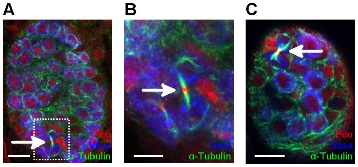
Figure 5. Feo is expressed in the larval germ cells.

Localization of Feo in larval gonads. First-stage larval testes (A,B) and ovaries (C) were stained with anti-Feo (red), anti-α-Tubulin (green) and anti-Vasa (blue) antibodies. Arrows indicate the localization of Feo at the spindle midbody in dividing germ cells. (B) Enlargement of the boxed area in (A). Scale bar represents 10 µm in H,J and 5 µm in I.
