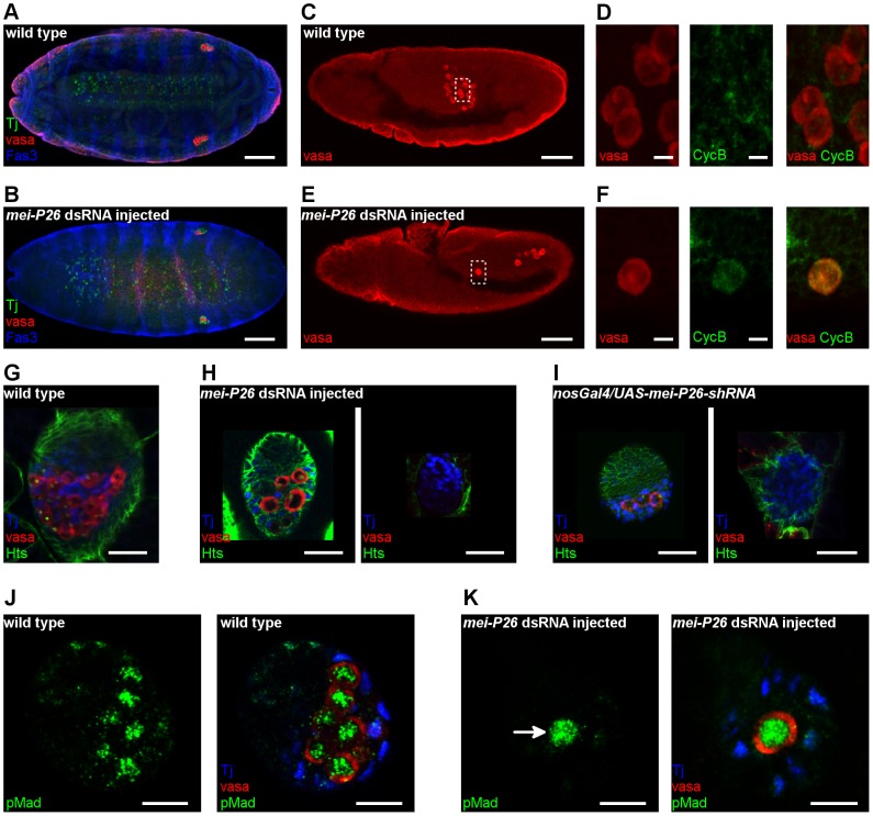
Figure 7. Mei-P26 regulates PGC development.
(A,B) Immunofluorescence images of embryos at stage 16, stained with anti-Vasa (red), anti-Tj (green) and anti-Fas3 (blue) antibodies. (A) Wild-type embryo. (B) Embryo injected with mei-P26 dsRNA has a reduced number of PGCs in the gonads. Ventral view is shown with anterior to the left. Scale bars represent 50 µm. (C–E) Immunostaining of embryos at stage 11 stained with anti-Vasa (red) and anti-CycB antibodies (green). Lateral view is shown; scale bars represent 50 µm in C,E and 5 µm in D,F. (C) Wild-type control embryos. (D) Enlargement of the boxed region in (C). In the wild-type embryos, germ cells do not express CycB. (E) Embryo injected with mei-P26 dsRNA. (F) Enlargement of the boxed region in (E). In embryos injected with mei-P26 dsRNA, germ cells express CycB prematurely. (G–I) Immunofluorescence images of ovaries of third-stage larvae stained with anti-vasa (red), anti-Hts (green) and anti-Tj (blue) antibodies. Scale bars represent 20 µm. (G) Wild-type ovary. (H,I) Larval ovaries with reduced mei-P26 function have rudimentary ovaries and contain few or no germ cells.(H) Ovaries of larvae treated with mei-P26 dsRNA. (I) Ovaries of nos-Gal4-VP16/UAS-mei-P26-shRNA larvae. (J-K) Immunostaining of ovaries at third larval stage with anti-Vasa (red) and anti-pSmad antibodies (green). DAPI labels the nuclei (blue). (J) Wild-type ovary. (K) Larval ovary treated with mei-P26 dsRNA. PGCs with reduced mei-P26 function express the Dpp downstream transducer pMad (arrow). Scale bars represent 10 µm.

