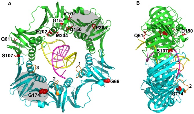
Figure 1. Summary of the positions of β clamp mutations.
Shown are (A) front and (B) side views of the β clamp on DNA (PDB: 3BEP). Amino acid positions bearing substitutions that failed to confer cold sensitive growth when co-overexpressed with Pol V are represented as red sticks in the green clamp protomer. The two residues mutated in the dnaN159(Ts) allele (β159; G66→E and G174→A) are indicated as red spacefill in the blue clamp protomer. Loops 1–3 of clamp are higlighted in orange in the blue clamp protomer; loops 1 and 2 contacted DNA in the crystal [5], [6]. The grey ovals represent the approximate location of the hydrophobic cleft present in each clamp protomer that contacts the CBM located in most, if not all clamp partners. This image was generated using PyMOL v1.5.0.2.

