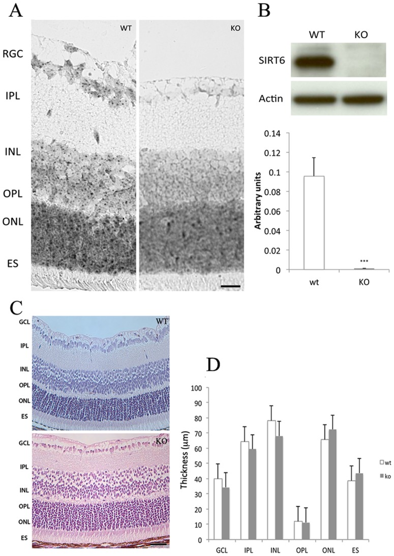
Figure 1. SIRT6 is expressed in the mouse retina.
A) Representative In situ hybridization showing the expression pattern of SIRT6 in 14-days-old mice (n = 3). Scale bar, 100 µm. B) Chromatin preparations from WT and KO mice retinas were analyzed by Western blot. β-actin was used as loading control (n = 4). Intensity of bands was determined by using the ImageJ software and is represented as arbitrary units. ***p<0.001 C) Retinal histological examination. Representative photographs from H&E stained retinal sections. 20X, scale bar 50 µm from WT and KO. No evident alterations in retinal morphologic features were observed. D) Quantitative analysis of retinal layers thickness. Ganglion Cell Layer (GCL), Inner Plexiform Layer (IPL), Inner nuclear Layer (INL) Outer Plexiform Layer (OPL), Outer Nuclear Layer (ONL), Retinal Pigment Epithelium (RPE). Data are the mean SEM (n = 4). NS, not significant.

