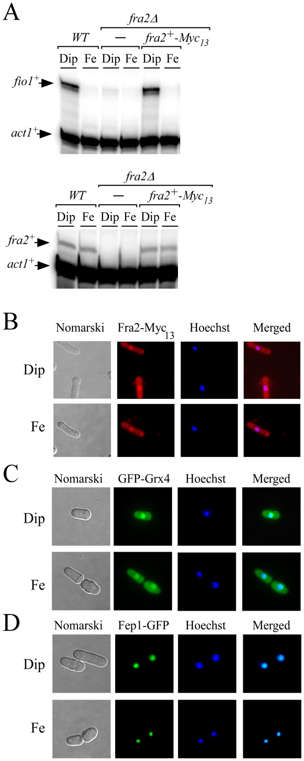
Figure 5. Fra2, Grx4, and Fep1 co-localize in the nucleus.
A, fio1+, fra2+ and act1+ mRNA steady-state levels were determined in a wild-type (WT) and a fra2Δ disrupted strain in which either an empty vector alone (-) or a wild-type copy of the fra2+-Myc13 allele was returned. Results are representative of three independent experiments. B-D, Cells expressing Myc13-tagged Fra2 (panel B), GFP-tagged Grx4 and Fep1 proteins (panels C and D) were treated with Dip (250 µM) or FeCl3 (Fe, 100 µM) for 30 min. Cells were analyzed by fluorescence microscopy for the presence of GFP (center left) and Hoechst stain (center right). Merged images are shown in the far right panels, whereas Nomarski pictures are depicted in the far left panels.

