Figure 1.

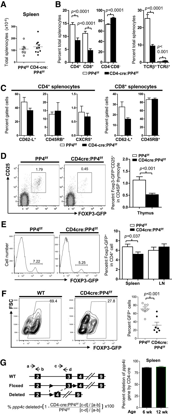
The ablation of PP4 induces partial αβ T lymphopenia and hampers Treg differentiation in vivo and in vitro. A, Splenocytes from 4-6 wk old mice were enumerated (n = 6-10). B-C, splenocytes were stained for various T cell markers, followed by flow cytometry analyses for the composition of CD4+, CD8+, αβ T, γδ T or CD4-CD8- cells in total splenocytes (n = 7-12) (B); alternatively, the expression of various lineage or activation markers on gated CD4+ or CD8+ cells was analyzed (n = 3-7) (C). D, Thymocytes were analyzed for the percentages of Foxp3-GFP+ cells in gated CD4SP population. Representative flow cytometry results are shown (left panels). Statistical analyses results are shown (right panel; n = 8 ~ 12). E, CD4-gated splenocytes were analyzed as in A. Representative flow cytometry results are shown (left panels). Statistical analyses results are shown (Right panel; n = 14 ~ 19). F, Naïve CD4+CD62-L+ cells were MACS-purified and activated under Treg polarization condition. Cells were harvested on d 3 to analyze Foxp3-GFP expression in CD4-gated population. Representative flow cytometry results and statistical analyses are shown (E = 4, n = 7). G, Splenic CD4+Foxp3-GFP+ Treg cells were sorted from 6 or 12 wk old mice. DNA from these cells was analyzed by qPCR to measure the extents of ppp4c gene deletion (see schematics for primer design and deletion efficiency calculation; left panel). Statistical analyses results are shown (right panel; n = 2). See Additional file 1: Figure S1 for flow cytometry gating strategies.
