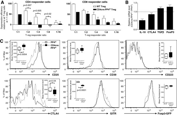
Figure 2.

PP4 deficiency impairs Treg suppression activity. A, CD4+Foxp3-GFP+ Treg cells (CD45.1-CD90.1-) were purified by sorting and co-cultured at titrating ratios with fixed numbers of CFSE-labeled WT responder CD4 and CD8 T cells (CD45.1-CD90.1+) and irradiated WT APC (CD45.1+CD90.1-). Division index of CD90.1+ CD4 and CD8 responder T cells was calculated from their CFSE patterns on d 3. Treg-mediated suppression was calculated by the division index differences between Treg-added samples and responder-only control. Relative suppression efficiencies were plotted after normalized to that of WT Treg cells at 1:1 ratio (E = 3, n = 4-5 group except for CD4cre:PP4f/f Treg at 1:1 ratio, for which n = 1 due to extremely low number of Foxp3-GFP+ cells in the CD4cre:PP4f/f mice). B, LN CD4+Foxp3-GFP+ Treg cells were sorted by flow cytometry. cDNA was synthesized using RNA from these cells and used for qPCR analyses. Relative mRNA level normalized to β actin results are shown (n = 2). C, LN CD4+Foxp3-GFP+ Treg cells were analyzed for various Treg markers and Foxp3-GFP expression. Representative plots on gated CD4+Foxp3-GFP+ cells are shown. Statistical analyses of the mean fluorescence levels are also shown (inserts; n = 8 ~ 10). See Additional file 1: Figure S1 for flow cytometry gating strategies.
