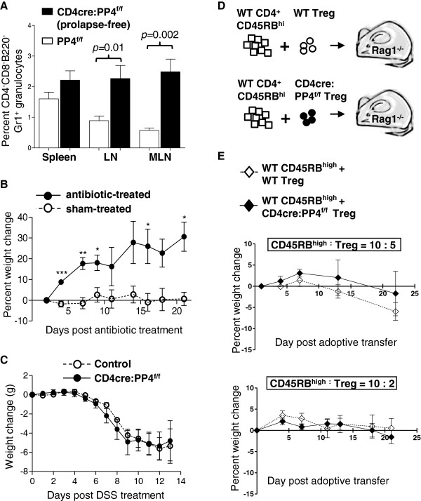
Figure 7.

Analyses of factors that may contribute to the onset of colitis in the CD4cre:PP4f/f mice. A, Cells from various tissues were stained and analyzed for lymphocyte marker expression. Percentages of CD4-CD8-B220-Gr1+ granulocytes in total cells are shown (n = 9-12). B, Prolapsed CD4cre:PP4f/f mice were treated with antibiotic in drinking water (antibiotic-treated) or left untreated (sham-treated). Percent weight changes of these mice up to 21 d are shown (E = 2, n = 3). C, Control or prolapse-free CD4cre:PP4f/f littermates were treated with 2% DSS in drinking water and monitored daily for weight changes for 13 d. D-E, WT CD4+CD45RBhigh cells (4x10⁵/mouse) were purified and co-transferred with sorted WT- or CD4cre:PP4f/f-Foxp3-GFP+ Treg cells (2x10⁵/mouse, top panel, n = 4; 8x10⁴/mouse, bottom panel, n = 10) into RAG1-/- mice (D). Recipients’ weight changes were monitored for 21 d (E). See Additional file 1: Figure S1 for all flow cytometry gating strategies.
