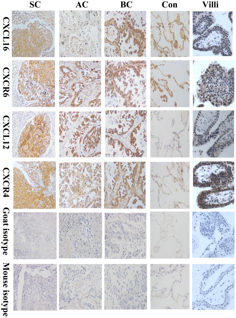
Figure 1. CXCL16-CXCR6 and CXCL12-CXCR4 were co-expressed in human lung cancer.
Immunohistochemistry was performed to determine the protein expression of CXCL16-CXCR6 and CXCL12-CXCR4 in human primary lung cancer tissues. The antibodies used in this experiments were mouse anti-human CXCR4 (25 µg/ml), mouse anti-human CXCR6 (25 µg/ml), mouse anti-human CXCL12 (20 µg/ml) and goat anti-human CXCL16 (20 µg/ml) antibodies. It was demonstrated in Fig. 1 that a specific brown-coloured staining for CXCL16-CXCR6 and CXCL12-CXCR4 in the cytoplasm and membrane of human different pathological types of lung cancer cells. Moderate-to-strong, brown-coloured staining for CXCL16 and CXCR6 was also observed in normal lung tissues, but the positive expression was mainly restricted to the alveolar epithelial cells and inflammatory cells. There was no evidence for nonspecific staining with the control antibody. The pictures were the representative of the experiments. AC: adenocarcinomas; SC: squamous carcinomas; BC: bronchoalveolar carcinoma; Con: normal lung tissues; Villi: human first-trimester villous tissues, as a positive control. Magnification, ×200.

