
Figure 4. The secretion of CXCL16 by lung cancer cell lines in vitro.
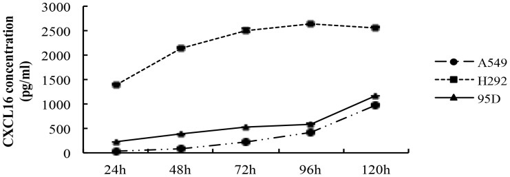
Digested A549, H292 and 95D cells were seeded in 24-well plates (500 µl/well) at a density of 5×10⁵/ml, respectively. Supernatants of the cell cultures were collected at 24, 36, 48, 60, 72, 96 and 100 h of culture. An ELISA assay was performed to examine the release of the soluble CXCL16 in cultured A549, H292 and 95D cells in vitro. As shown in Fig. 4, three kinds of lung cancer cell lines all secreted CXCL16 spontaneously in a time-dependent manner, despites of a difference in the concentration of CXCL16 in the culture medium. The experiments were repeated three times. Error bars depict the standard error of the mean.