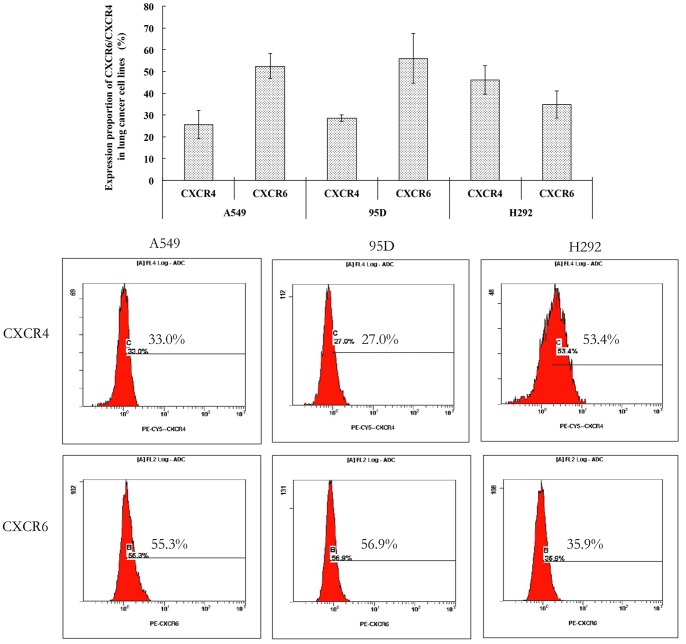
Figure 5. The membrane expression of CXCR6 and CXCR4 in lung cancer cell lines in vitro.
Flow cytometry (FCM) was used to detect the membrane expression of CXCR6 in lung cancer cell lines. The cells of 1×10⁵ were counted and the dilution ratio of (phycoerythrin)-CXCR6 monoclonal antibody and PE-CY5-CXCR4 monoclonal antibody were 1∶10 and 1∶5, respectively. The histogram demonstrated the average expression proportion of CXCR6 and CXCR4 in A549, H292 and 95D cells, respectively. The FCM pictures were representative of the experiments. The percentage of membrane CXCR6-positive cells in A549, H292 and 95D was 52.4±5.80, 56.03±11.42 and 34.8±6.17, respectively. Furthermore, the membrane expression of CXCR4 was also observed in A549, H292 and 95D cells at the same time. The experiments were repeated three times and the images were representative of the experiments. Error bars depict the standard error of the mean.

