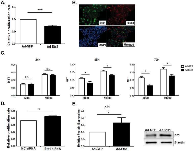
Figure 5. Ets1 inhibits Min6 cell proliferation.
(A). Min6 cells were infected with Ad-EGFP or Ad-Ets1 for 24 hrs and cell proliferation was determined by EdU labeling. (B). Min6 cells were infected with Ad-Ets1 and incubated with BrdU. Overexpressed Ets1 was detected with rabbit anti-Ets1 antibody and BrdU labeling with mouse anti-BrdU antibody. (C). Min6 cell proliferation was also determined by MTT assay after 24, 48 or 72 hrs post-infection. (D). Min6 cells were transfected with Ets1 siRNA and cell proliferation was determined by EdU labeling. Ets1 siRNA treatment increased Min6 cell proliferation as shown by EdU labeling. (E). Ets1 induced p21 expression in Min6 cells. Min6 cells were infected with Ad-Ets1 and p21 protein levels were detected by Western blot. (n = 3; values are shown as mean ± SD. *p<0.05, **p<0.01, ***p<0.001. N.S., not significant, t-test).

