
Figure 2.
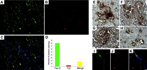
Tau oligomers constitute a small portion of total tau in AD frontal cortex. A–C) Representative immunofluorescent images of AD frontal cortex showing Tau-5 (A, green), T22 (B, red), and merge (C; also including DAPI, blue) confirm the presence of tau oligomers in situ. D) Histogram summarizing quantitation of 16 AD cases; only 20% of total aggregates corresponded to oligomers. Extracellular tau oligomers were also detected. E–H) Photomicrographs demonstrating diffuse extracellular tau oligomers (as detected by T22), including those in association with arterioles. Double immunofluorescence with von Willebrand factor antibody (I, green), T22 (J, red), and merge (K; also with DAPI, blue) suggested the presences of extracellular tau oligomers. Sections were stained with DAPI. Scale bar = 20 μm (A–C); 15 μm (E–K).