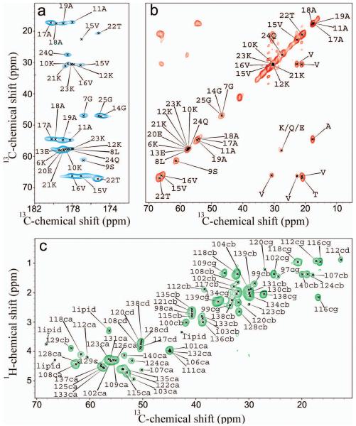
Figure 1. MAS ssNMR spectrum of αS bound to DOPE:DOPS:DOPC SUVs.
13C-13C DARR correlation spectrum recorded at −19 °C using a 20 ms contact time at a MAS rate of 10 kHz. Carbonyl and aliphatic regions are showed in panels A and B, respectively. Residue names are reported using the single letter convention. c) 1H-13C correlation via INEPT transfer recorded at 4 °C at a MAS rate of 10 kHz. The experiments were performed at 1H frequencies of 600 and 700 MHz using a 1H/13C 3.2-mm probe and a spinning speed of 10.0 kHz. Atom names ca, cb, cg, cd are used for Cα, Cβ, Cγ and Cδ atoms, respectively.

