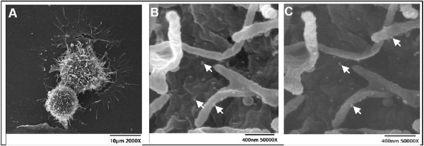
Figure 4.

MUC16 on the surface of ovarian cancer cells. OVCAR-3 cells were labeled with VK8 followed by colloidal gold nanoparticles conjugated with goat anti-mouse secondary antibody. A, low magnification secondary electron image of two labeled OVCAR-3 cells. B, Scanning Electron Microscopy (SEM) image of OVCAR-3 showing colloidal gold nanoparticles binding to cell surface and microvilli. C, Back scattered electron image of same cell surface shown in B, clearly showing the colloidal gold nanoparticles. Bright spots (some indicated by bright arrows) in B and C are the colloidal gold nanoparticles. OVCAR-3 cells are not labeled with colloidal gold nanoparticles in the absence of VK-8 (data not shown).
