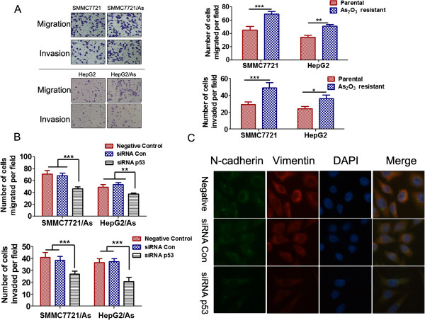
Figure 3.

Mutp53 contributed to the increased metastatic potential of HCC resistant cells. (A) The migration and invasive abilities of HCC parental and arsenic trioxide resistant cells were determined by trans-well assays in chambers coated with matrigel (for invasion assays) or without matrigel (for migration assays). Left panels: representative images; right panels: quantifications of average number of cells/field. *P < 0.05, **P < 0.01, ***P < 0.001, two-way ANOVA with Bonferroni post-test. (B) Results of the migration and invasion assays for the HepG2/As and SMMC7721/As cells transfected with p53 siRNA or control siRNA. (**P < 0.01, ***P < 0.001, two-way ANOVA with Bonferroni post-test). (C) Single and merged images were taken to show immunofluorescence staining of N-cadherin (green) and vimentin (red) accompanied by the cell nucleus (blue) stained by DAPI.
