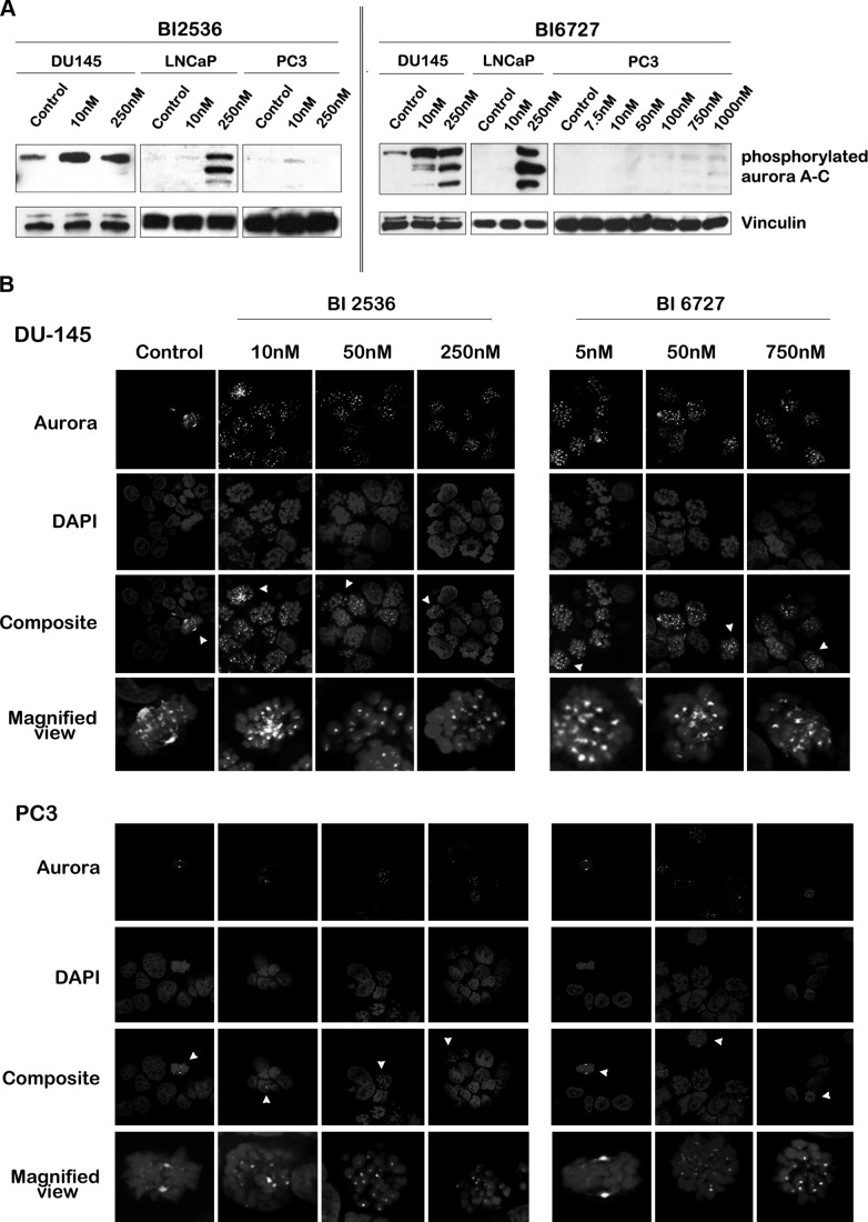
Figure 4.
Expression and localization of active aurora kinases differ between PCa cells after treatment with BI 2536 and BI 6727. A) Western blot analyses for phosphorylated aurora A–C (from high to low molecular weight: aurora A, B, C) were performed after 24 h of Plk1 inhibition with BI 2536 and BI 6727 in PCa cell lines. Protein expression levels of phosphorylated aurora were increased in DU145 and LNCaP cells after BI treatment, but not in PC3 cells. B) DU145 (top panels) and PC3 (bottom panels) cells were treated for 24 h with BI 2536 (left panels) or BI 6727 (right panels), fixed and stained with DAPI, and aurora antibody. Cells were visualized under a confocal microscope. A representative cell (arrowhead) is magnified below. Phosphorylated aurora localized to microtubules and mitotic poles of control cells undergoing mitosis. Cells treated with BI 2536 or BI 6727 lacked polar aurora staining but exhibited aurora at the kinetochores. Image is representative of ≥3 independent experiments.

