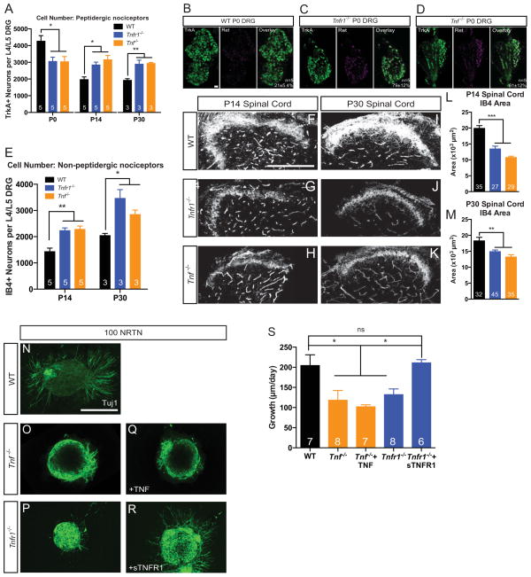
Figure 4. Deletion of Tnfr1 or Tnf leads to the premature differentiation and impaired growth of non-peptidergic nociceptors.
(A) Quantification of the number of TrkA+ sensory neurons in the L4/L5 DRG at P0, P14, and P30.
(B–D) Representative images of Ret/TrkA colocalization in the P0 DRG from WT, Tnfr1−/−, and Tnf −/− mice. Scale bar represents 30 μm. Quantification of colocalization shown on overlay.
(E) Quantification of the number of IB4+ neurons in the L4/L5 DRG at P14 and P30.
(F–M) Immunostaining and quantification of IB4+ axons invading the spinal cord dorsal horn at P14 (F–H) and P30 (I–K) in WT, Tnfr1−/−, and Tnf −/− mice. Quantification of IB4+ axon area shown in (L–M). Scale bar represents 100 μm.
(N–S) E14.5 DRG explants cultured in 100 ng/mL of NRTN (N–P), 100 ng/mL NRTN + 2 ng/mL TNF (Q), or 100 ng/mL NRTN + 5 μg/mL sTNFR1 (R) for 24 hours. Explants from ≥3 mice per condition. Scale bar represents 100 μm.
1-way ANOVA, Bonferroni post-test (A,L,M) or Tukey post-test (S). 2-way ANOVA, Bonferroni post-test (E). Number of mice analyzed is indicated in A,E. 5 mice analyzed in L–M.
Data represent mean±SEM, ns: not significant, *p<0.05, **p<0.01, ***p<0.001.

