
Figure 6. Pax5 expression in young and adult spleen tissue.
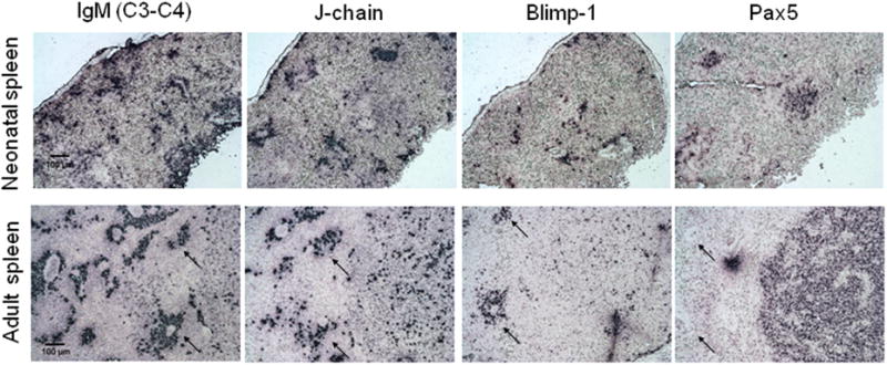
In situ hybridization staining of 8μm (non consecutive) sections of spleen tissue from a 1 month old (shark H) (A) or adult shark (shark W) (B). From left to right probes are specific for: IgM spanning C3-C4, J-chain, Blimp- or Pax5. Black arrows indicate regions near blood vessels where plasma cells congregate. All photographs taken at 10× magnification with 100 μm scale bar shown. Positive staining is purple. Negative controls with sense probes showed no staining (not shown).