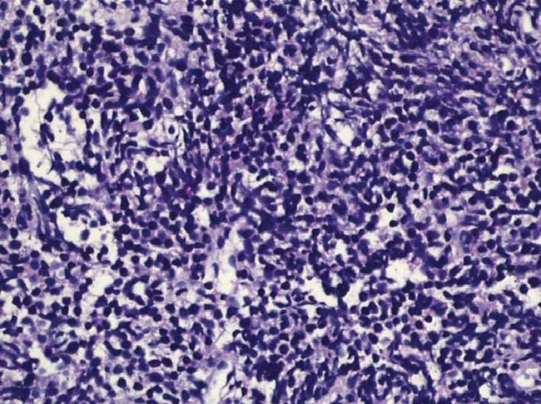
Figure 2.

Small cell carcinoma of the prostate formed by malignant epithelial cells with narrow cytoplasm in the fibromuscular stroma. This figure shows us the microscopic findings of small cell carcinoma of the prostate in the pathologic examination. The specimen was obtained after a transrectal ultrasound-guided prostate biopsy of the patient.
