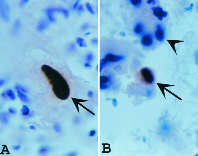
FIG. 1.
(A) Typical positive immunohistochemistry (IHC) staining for CMV (arrow) in a cytomegalic cell; (B) the type of cell designated atypical in this study (arrow); it was positive for CMV by IHC but not cytomegalic. The cells that stain positive for the presence of CMV (arrows) are a distinctive brown, whereas the cells that are negative for CMV by IHC (arrowhead) are blue, secondary to counterstaining with H&E. Magnification ×1,000.

