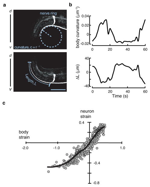
Figure 1. The shape of the touch receptor neuron AVM as a function of stress evoked by body movement.
a, Representative micrographs of a GFP-labeled AVM neuron in an adult worm bending ventrally (top) or dorsally (bottom). Curvature and neuron length are schematically indicated as dashed lines. Scale bar = 50 μm.
b, Body curvature (top) and the change in neuron length (bottom) vs. time. Epochs of ventral bending correspond to positive curvature and AVM shortening, while epochs of dorsal bending correspond to negative curvature and AVM extension.
c, Estimated neuron strain, ΔL/L as a function of body strain. Body strain was derived by approximation of the worm as an Euler-Bernoulli tube49, as described in Supplementary Note 1. The black line is a smoothed version of the neuron strain-body strain relationship. Data drawn from n=7 control, TRN::GFP transgenic worms and 352 still images collected during four independent imaging sessions.

