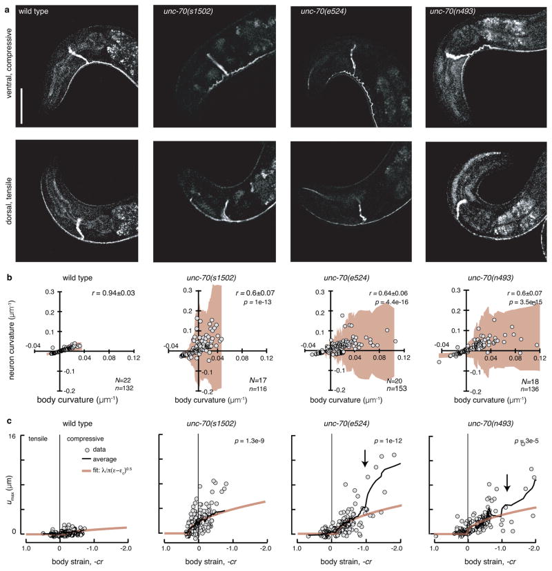
Figure 2. Loss of unc-70 β-spectrin function causes buckling in TRNs during ventral bending.
a, AVM shape during ventral (top) and dorsal (bottom) bending in wild-type and unc-70 mutant TRN::GFP animals. Scale bar is 50 μm.
b, Neuron curvature vs. body curvature. Each point is the mean of 10–30 adjacent curvature measurements in a still image (see Methods). Shaded areas (beige) indicate the standard deviation of neuron curvature; the increased deviation during compressive (positive, ventral) body bends indicates a larger variation in local neuron curvature. r is the correlation coefficient between neuron and body curvature and r values in unc-70 mutants were significantly different than wild-type (Fisher z-transform of r). p values indicated in the upper right. N=Number of animals and n=number of still images. Result is from three independent imaging sessions.
c, Maximum off-axis deformation (buckling), umax, of AVM as a function of body strain. Each point is the maximum deformation within a still image. Black lines are running averages of 15 images; beige lines are the calculated buckling deformation of a constrained, flexible filament bundle as a function of compressive strain ε (see Supplementary Note 1). p values derived from linear regression and compared to wild-type indicated in the upper left. Same numbers of observations as in b. All strains carry the uIs31 transgene encoding TRN::GFP.

