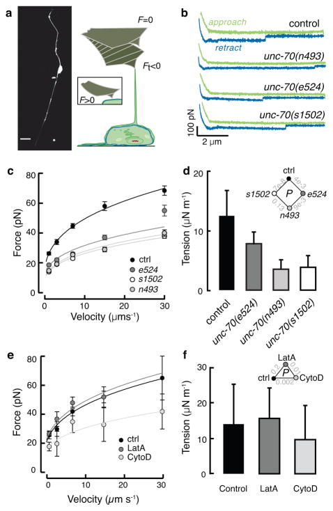
Figure 3. Loss of β spectrin function decreases TRN tension.
a, Micrograph of a GFP-tagged TRN in vitro (left) and schematic of the tether extrusion procedure. A peanut lectin-coated AFM cantilever was held in contact with each cell for about 400 ms and a lipid tube formed following retraction in about 20% of cases; such events result in a downward deflection of the cantilever (Ft < 0).
b, Representative force-distance curves (approach in green, withdrawal in blue) acquired at 7 μm·s−1. The discontinuity during withdrawal indicates an interaction event and is proportional to the membrane tension. Similar results were observed in more than 17 cells/genotype.
c, Tether force (mean±SEM) vs. cantilever retraction velocity in control and mutant TRNs. Force-velocity curves were fit to a power law and used to estimate the static tether force at zero velocity (see Methods). Supplementary Table 3 lists the number of cells and tethers for each condition. Data in b and c were acquired during n=6 independent AFM sessions.
d, The apparent membrane tension, Tapp, of cultured TRNs, derived from the static tether force, fo, estimated from the fits to the data in c according to: Tapp = fo2/8ππ2κ and a value for the membrane bending rigidity, κ of 2.7·10−19 N·m (Ref. 29). Diamond inset shows p values for the tested combinations.
e, Tether force (mean±SEM) vs. cantilever retraction velocity in the presence and absence of drugs that disrupt actin. LatrunculinA (LatA) and cytochalasin D (cytoD) applied at 1 and 2 μM, respectively. More than 25 tethers were tested for each speed, except for 30μm/s since no data were obtained from LatA-treated cells under this condition. Supplementary Table 3 lists the number of cells and tethers for each condition. Treated and untreated cells were tested in parallel during n=3 independent AFM sessions.
f, Membrane tension is unaffected by actin depolymerizing drugs. Triangle inset shows p values as a function of treatment. P-values derived after log-log transformation of the force-velocity data and linear regression followed by a Tukey-type t-test. All strains carry the uIs31 transgene encoding TRN::GFP, which was used to identify TRNs in cultures.

