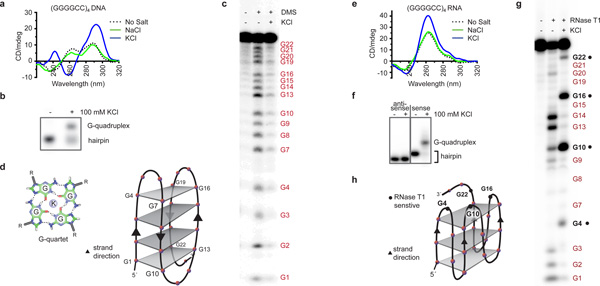
Figure 1. DNA and RNA of the C9orf72 HRE form G-quadruplexes.
a) The CD absorptivity shows characteristic K+-dependent spectra for antiparallel DNA G-quadruplexes with (GGGGCC)4. b) The presence of K+ during annealing induces a conformational change that decreases the mobility of (GGGGCC)4 DNA in a gel mobility shift assay. c) A DMS footprinting assay on (GGGGCC)4 DNA shows protection of the N7 positions on all the guanines when a G-quadruplex is formed. d) The proposed topology for the antiparallel DNA G-quadruplex formed by (GGGGCC)4. Each gray plane represents a G-quartet, as shown in the lower corner. Four separate G-quartets are stacked 5´ to 3´ with two cytosines forming each loop region. e) The CD spectra identified the formation of a parallel G-quadruplex for the RNA (GGGGCC)4 in the presence of 100 mM KCl. f) (GGGGCC)4 RNA demonstrates a slower mobility in the presence of KCl compared to (CCCCGG)4 RNA. g) The RNase T1 protection assay identifies single-stranded guanine residues (denoted in black) not involved in the formation of the G-quadruplex. h) The proposed parallel G-quadruplex topology formed by the RNA of the C9orf72 HRE. Three stacks form the parallel G-quadruplex, with the RNase T1-sensitive guanines shown as black dots.

