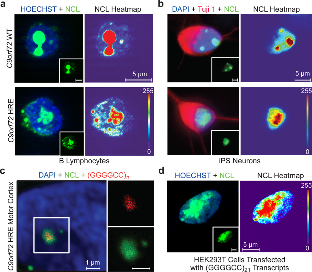
Figure 4. Nucleolar stress is a result of repeat-containing RNA transcripts from the C9orf72 HRE.
a) In the control C9orf72 WT B lymphocytes, NCL (green) is localized to the condensed nucleolus. In contrast, the cells of patients with the C9orf72 HRE show an increased NCL diffusion and fractured nucleoli in the nucleus (Hoechst, blue). A heat map of NCL intensities marks the difference between cells. b) iPS motor neurons derived from patients carrying the C9orf72 HRE also demonstrate NCL mislocalization. β-III tubulin (Tuj1) (red) was used to identify neurons. c) NCL colocalizes with RNA foci (red) formed in motor cortex tissue from patients carrying the C9orf72 HRE. A (CCCCGG)2.5 probe was used to detect the (GGGGCC)n RNA foci, a previously identified pathological feature of the C9orf72 HRE tissues. d) Transfection of (GGGGCC)21 abortive transcripts (Figure 2a) recapitulates NCL pathological features observed in patients cells with the C9orf72 HRE.

