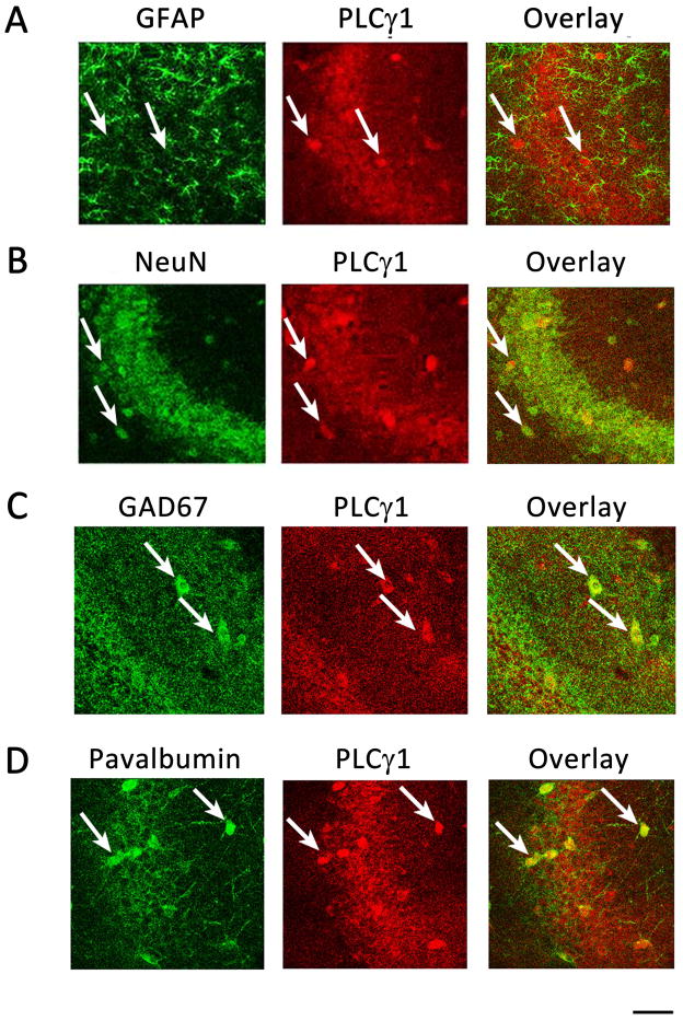
Figure 4. PLCγ1-immunoreactivity is localized in hippocampal neurons.
(A, B) PLCγ1-ir is localized in hippocampal principal neurons. Representative images in high magnification of CA3a. PLCγ1-ir is localized to NeuN-ir+ (B) but not GFAP-ir+ (A) cells. (C, D) PLCγ1-ir is also localized in hippocampal interneurons. (C) Representative images from CA1 stratum radiatum with PLCγ1 (red) and GAD67 (green). PLCγ1-ir colocalized with GAD67-stained cells (arrows). (D) Representative images from CA3a, showing most PLCγ1-ir+ cells are Parvalbumin-ir+ (arrows). Scale bar: 50 μm.

