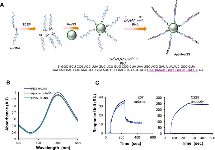
Figure 1.
(A) Schema for the conjugation of aptamer to HAuNS. (B) Absorbance spectrum of apt-HAuNS, C225-HAuNS, and PEG-HAuNS in water, which peaked at 800 nm. (C) Representative surface plasmon resonance sensorgrams of aptamer and C225 antibody on sensor chips coated with rhEGFR. Each ligand was injected and analyzed in duplicate binding cycles. The vertical axes in response units represent binding of each ligand to immobilized rhEGFR.

