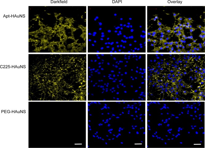
Figure 3.
Selective binding of aptamer-coated HAuNS to OSC-19 cells. Only cells incubated with aptamer- or C225-coated HAuNS had a strong light-scattering signal. Cells were stained with DAPI for visualization of cell nuclei (blue). Light-scattering images of HAuNS were pseudocolored yellow. Bar: 50 μm.

