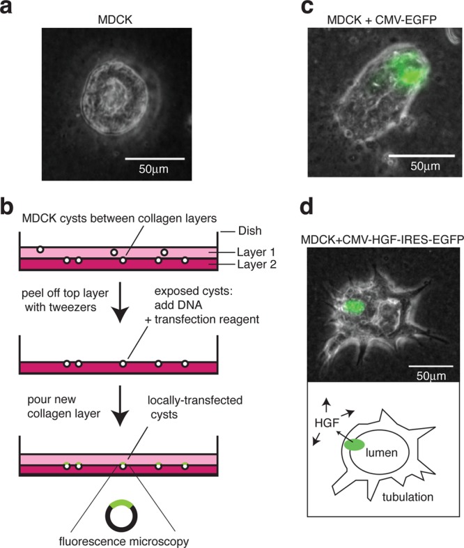
Figure 1.

Localized transfection method for 3D MDCK culture. (a) MDCK cells seeded into collagen grow into hollow spherical cysts (∼200 cells) over a period of 6 days. (b) Schematic view showing two collagen layers in a 35 mm diameter dish. A second layer, comprising collagen mixed with the seeding cells, allows cysts to develop at the interface between the layers. The top layer is peeled away to allow transfection of one face of the cysts. A new collagen layer is then poured on top. (c) A typical MDCK cyst, locally transfected with EGFP under a constitutive CMV promoter. (d) Local transfection with HGF under a CMV promoter, followed by an internal ribosome entry site (IRES) and the EGFP gene. A schematic view (below) shows the transfected region (green) secreting functional HGF, which diffuses around the cyst to induce tubulation. Phase contrast light microscopy images are superimposed with the GFP fluorescence channel. Scale bars: 50 μm.
