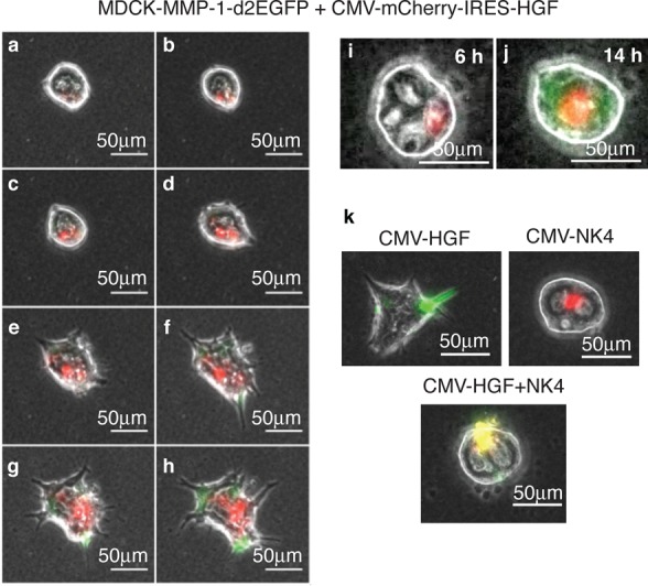
Figure 5.

Localized sender–receiver patterns in single MDCK cysts. The MDCK-MMP-1-d2EGFP reporter cyst is locally transfected with pCMV-mCherry-IRES-HGF (red region). The red region secretes HGF which diffuses extracellularly to induce distal GFP expression and tubule formation. (a–h) Images taken from 6 h after transfection, at 2 h intervals. A typical cyst is shown and can also be seen in timelapse (Supporting Information Movie S2). (i, j) An independent example of the same setup at 6 h (i) and 14 h (j); this cyst goes on to tubulate after 14 h (Supporting Information Movie S3). (k) Locally transfecting MDCK wt cysts with CMV-EGFP-IRES-HGF activator (green) or mCherry-IRES-NK4 (red), or both together (yellow). Images taken after 24 h. Cotransfecting NK4 blocks HGF-induced tubulation. Scale bars, 50 μm.
