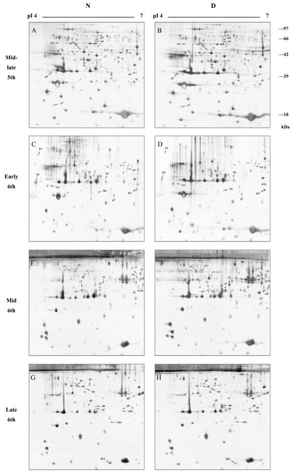
Figure 1.

Representative 2-DE images of the proteins from H. armigera larval hemolymph. Proteins were extracted from nondiapause- (N) and diapause-destined (D) larval hemolymph at four stages: (A and B) mid-late stage of the fifth instar, (C and D) early stage of the sixth instar, (E and F) middle stage of the sixth instar, and (G and H) late stage of the sixth instar. Proteins were separated by IEF (18 cm IPG strips, pH 4–7, L) and 12% SDS-PAGE. Gels were stained by MS-compatible silver stain. Based on triplicate replications, only statistically significant (p-value < 0.05) protein spots that changed ≥1.5-fold were considered for further analysis. Differentially expressed protein spots are marked with letters and numbers. Letters 'a’, 'b’, 'c’, and 'd’ represent the mid-late stage of the fifth instar, the early, the middle, and the late stages of the sixth instar, respectively.
