
Figure 2.
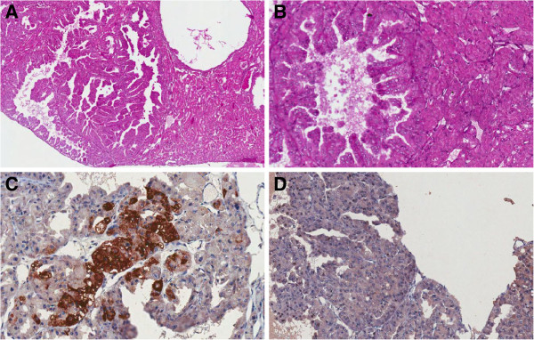
Histological section of a papillary neoplastic proliferation of a kidney from an (AIRmax x AIRmin)F2 mouse. Panel A, hematoxylin and eosin stained section, 40X magnification. Panel B, same section at 250X magnification. The renal tumor resulted positive for racemase (AMACR) immunostaining (panel C, 250X magnification) and negative for CD10 immunostaining (panel D, 250X magnification).