
Figure 2.
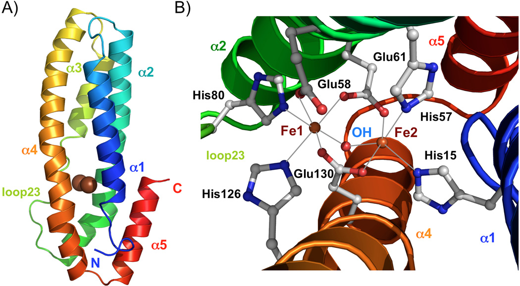
Structure of oxidized FBXL5 hemerythrin-like domain. A) Ribbon representation of the FBXL5 Hr structure. The two iron atoms are shown with the brown spheres. B) Close up of the diiron center. Iron ligands of the irons are shown in ball-and-stick representation. The red sphere respresents the μ-hydroxo bridge.