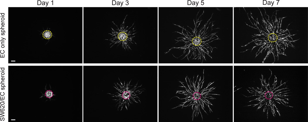
Fig. 2.
Development of EC only spheroids and SW620/EC spheroids over 7 days in a fibrin gel. EC only spheroids exhibit robust sprouting angiogenesis (top panel). When combined with SW620, EC initially reorganize to periphery of the spheroids, then infiltrate to form inner network within the boundaries of the spheroid that is contiguous with outer sprouting vessels (bottom panel). EC are transduced with EGFP (white). Dashed lines outline original spheroid boundaries from Day 1. Scale bars represent 500 µm.

