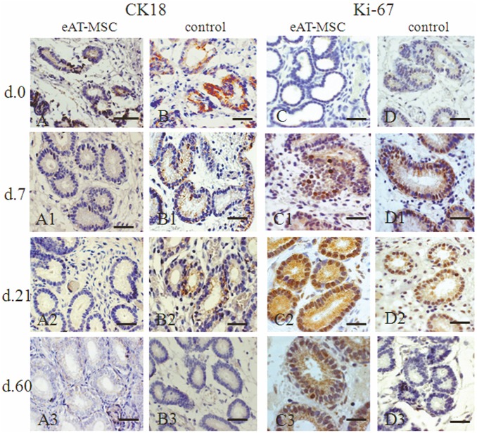
Figure 4. Cytokeratin 18 (CK18) and Ki-67 expression before (at day 0) and after (at days 7, 21 and 60) eAT-MSCs intrauterine transplantation: A–A3 and D–D3 - experimental group; B–B3 and D–D3 - control.
A, B) At day 0, CK18 (black arrows) localized in damaged epithelia of glands (G). A1–A3) At days 7, 21 and 60, the absence of CK18 expression was observed. B1, B2) At days 7 and 21, CK18 expression was still observed (black arrows) in control. B3) At day 60, control mares showed no signs of CK18 expression. C, D) At day 0, none or a few Ki-67 positive cells were observed. C1, D1) At day 7, amount of Ki-67 positive cells (black arrow) was increased. C2, D2) At day 21, both groups showed positive Ki-67 staining. C3) At day 60, the expression of Ki-67 was still observed. D3) In control, the absence of Ki-67 expression. LM. Scale bars: A–D1 = 50 µm; C2, C3, D2, D3 = 25 µm.

