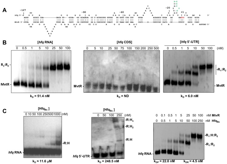
Figure 2. MtvR regulates hfq Ec expression through interaction with its 5′-leader region.
(A) Schematic representation of the nucleotide interaction between the 5′-UTR of hfq Ec and the MtvR sRNA, highlighting in green and red lettering, respectively, the RBS and the AUG translation start site in the hfq Ec 5′-UTR. (B) EMSA experiments using 25 nM of biotin-labeled MtvR transcript and the indicated concentrations of: left panel, the hfq Ec full mRNA; center panel, the hfq Ec coding region (CDS); right panel, the 5′-UTR of hfq Ec. C) EMSA experiments performed to assess the ability of HfqBc to bind to: left panel, 25 nM of the biotin-labeled hfq Ec full mRNA; center panel, 25 nM of the biotin-labeled hfq Ec 5′-UTR. The ability of mixtures containing HfqBc together with the tested concentrations of the MtvR transcript to bind to 25 nM of the biotin-labeled hfq Ec full mRNA is shown in the right panel. H(n), HfqBc(n); R1, MtvR; R2, hfq Ec RNA; kDP, affinity constant for the protein; kDR, affinity constant for the RNA species.

